
Fluorescent labeling: Principles, applications, and modern techniques
What is fluorescent labeling?
Fluorescent labeling is a technique that attaches fluorophores to biological molecules, enabling visualization in microscopy, flow cytometry detection or other methods and diagnostics.
- Learn more about Flow Cytometry here: Flow Cytometry Glossary
Chemically, fluorophores bind selectively and vary in excitation and emission wavelengths, while physically, fluorescence occurs through light absorption and emission, governed by the Stokes shift for optimal detection.
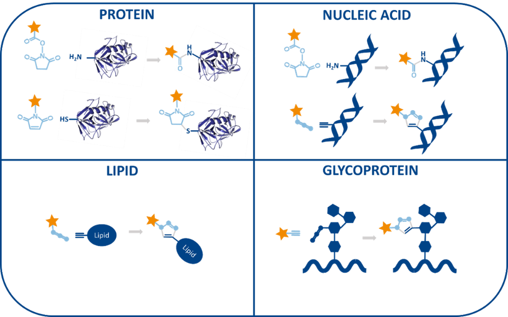
The fluorescent labeling process
The fluorescent labeling process involves target selection, fluorophore choice, chemical conjugation (amine, thiol, enzymatic, click chemistry, genetic tagging), purification, and validation.
Fluorophores can be intrinsic (naturally fluorescent) or extrinsic (synthetically attached). Common types include:
– Organic dyes – Small synthetic molecules like fluorescein, rhodamine, Alexa Fluor, used for antibody labeling and DNA probes.
– Fluorescent proteins – Genetically encoded proteins such as GFP, mCherry, CFP/YFP, ideal for live-cell imaging.
– Quantum dots – High-brightness nanocrystals for multiplex detection.
– Fluorescent nanoparticles – Silica beads, polymer dots for targeted imaging and drug delivery.
– Environment-sensitive dyes – Fura-2, BCECF, DiI, for functional biosensing.
Kit solutions for fluorescent labeling
Fluorescent labeling kits include multiple components like: fluorophores, conjugation reagents, buffers, blocking reagents, purification tools and protocols to ensure efficient labeling with high sensitivity and specificity.
Fluorescent labeling kits attach fluorophores to biomolecules through specific chemical reactions, ensuring precise detection in imaging and analytical applications. For proteins, they use NHS esters or maleimides to label lysine or cysteine residues, while nucleic acids can be labeled using fluorescent probes, intercalating dyes, click chemistry, or NHS esters, enabling applications such as FISH, qPCR, and molecular beacon probes. Lipids, small molecules, and carbohydrates are tagged using lipophilic dyes, click chemistry, or hydrazide-functionalized fluorophores, facilitating studies in membrane tracking, biosensing, and glycan profiling.
Kit fluorescent labeling for cytometry
Fluorescent labeling kits for flow cytometry are essential tools that enable precise, sensitive detection of specific cellular targets by tagging biomolecules with photostable fluorophores through robust conjugation methods. They are optimized for compatibility with flow cytometers, minimizing spectral overlap and background noise, while supporting multiplexed analysis for complex applications like immunophenotyping, apoptosis detection, and cell proliferation studies. Many kits also include blocking agents and compensation controls to improve signal clarity and reproducibility across experiments.
Kit fluorescent labeling for high throughput screening
High-throughput screening (HTS) plays a crucial role in modern drug discovery by enabling fast, automated testing of large compound libraries to uncover promising therapeutic candidates. It utilizes miniaturized assays, robotic automation, and advanced analytics to accelerate early development stages. With consistent reagents, multiplexed readouts, and biologically relevant models, HTS delivers high-quality data. Its scalability and integration with software tools make it a cost-effective solution for large-scale screening.
Kit fluorescent labeling for imaging
Kit fluorescent labeling for imaging are designed to streamline workflows, improve reproducibility, and enhance image quality across a wide range of biological and material science applications.
| Microscopy Technique | Kit Type | Applications | Key Optimization Features |
| Brightfield Microscopy | Histology, Gram, Cytology, PAS | General tissue morphology, bacterial ID, cell shape | Consistent staining, high contrast, reproducibility |
| Fluorescence Microscopy | IF Kits, Live-cell,
Organelle |
Protein localization, real time activity, subcellular imaging | Specific signal, low background, photostable dyes |
| Viability, Apoptosis/Necrosis | Live/dead cell detection, apoptosis study | Easy dual-stain, sensitive detection via flow/fluorescence | |
| Confocal Microscopy | 3D Cell Culture, Live Imaging Kits | 3D imaging, dynamic live cell tracking | High resolution, reduced background, phototoxicity control |
| Super-Resolution Microscopy | STED/SIM, Expansion, DNA-Paint | Nanoscale imaging, dNA hybridization, tissue expansion | High spatial resolution, multiplexed targeting, high photon yield |
| Electron microscopy (EM) | TEM/SEM prep, Immunogold, Stain | Ultrastructure & virus visualization, protein localization | High contrast, sharp detail, nanometer resolution |
| Multiphoton/Deep Tissue | Tissue clearing, Deep staining | 3D deep tissue imaging, thick tissue labeling | Organ-level imaging, long term signal stability |
Fluorescent labeling kits are widely used tools in cell biology and medical imaging, enabling the visualization of structures, molecules, and processes with high specificity and sensitivity and so widely applied in cell biology and medicinal imaging:
- Tracking Live cell proliferation, which is crucial in immunology and cancer research
- Cytoskeletal studies, which leads to visualize the cytoskeleton of fixed cells, helping in studies of cell shape, motility and structural changes during disease progression.
- Tracking protein trafficking and localization in live cells
- Tumor imaging using fluorescent antibody conjugates
- Blood-brain barrier permeability studies
Advantages and challenges in fluorescent labeling
Technical challenges in fluorescent labeling
Fluorescent labeling is a powerful technique, but it comes with a handful of technical hurdles that researchers often have to navigate:
- Low Signal & Poor Labeling: Inefficient or incomplete labeling leads to weak or inconsistent fluorescence.
- Background & Specificity Issues: Non-specific binding and autofluorescence reduce image clarity.
- Photobleaching: Fluorescent signals fade over time with light exposure, limiting long-term imaging.
- Toxicity: Some dyes and reagents can harm live cells.
- Spectral Overlap: Fluorophores may interfere with each other, making multiplexing difficult.
- Reproducibility & Complexity: Batch variations and complicated protocols can reduce reliability.
Benefits of fluorescent labeling
This fluorescent labeling imaging technique offers several advantages in biomedical research and diagnostics:
- High sensitivity allows detection of even trace levels of biomolecules.
- High specificity enables precise targeting using antibodies or molecular probes.
- Real-time imaging facilitates the observation of live-cell processes as they happen.
- Multiplexing lets researchers visualize multiple molecular targets simultaneously.
- Quantitative capabilities support accurate measurement of molecule abundance.
- Versatile target range includes DNA, RNA, proteins, lipids, and glycans
- Automation compatibility makes it suitable for high-throughput workflows.
- Gentle sample handling ensures minimal disruption thanks to biorthogonal tools.
- High-resolution imaging is essential for cutting-edge microscopy techniques.
- Clinical relevance stems from its widespread use in diagnostics and surgical imaging.
baseclick offers a wide range of fluorescent dyes for labeling biomolecules using mild and efficient click chemistry.