

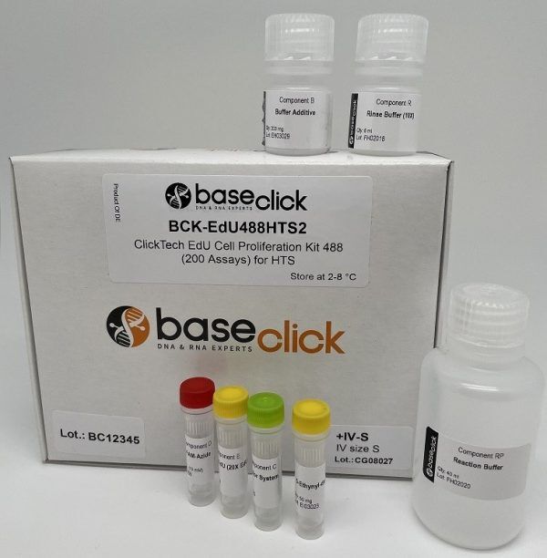
In Vivo EdU Cell Proliferation Assay for High-throughput Screening
ClickTech In Vivo EdU Cell Proliferation Kit for HTS
| Size | Catalog No. | Price |
|---|---|---|
| Dye 488 / 200 Assays / Size S | BCK-EdU488HTS2+IV-S | € 415,00 |
| Dye 488 / 200 Assays / Size M | BCK-EdU488HTS2+IV-M | € 550,00 |
| Dye 488 / 200 Assays / Size L | BCK-EdU488HTS2+IV-L | € 695,00 |
| Dye 555 / 200 Assays / Size S | BCK-EdU555HTS2+IV-S | € 415,00 |
| Dye 555 / 200 Assays / Size M | BCK-EdU555HTS2+IV-M | € 550,00 |
| Dye 555 / 200 Assays / Size L | BCK-EdU555HTS2+IV-L | € 695,00 |
Chemical Properties
-
Shelf Life
12 months unopened after receipt
-
Storage Conditions
2-8 °C
-
Physical State
kit system made of different components
-
CAS Number
n.a.
-
Excitation (max)
Dye 488: 496 nm | Dye 555: 546 nm
-
Emission (max)
Dye 488: 516 nm | Dye 555: 579 nm
-
Ɛ (max)
Dye 488: 83.000 cm-1M-1 | Dye 555: 91.000 cm-1M-1
-
Preparation/Handling
please see user manual of the kit
Product Information
Accurate quantification of DNA synthesis in vivo for high-throughput applications
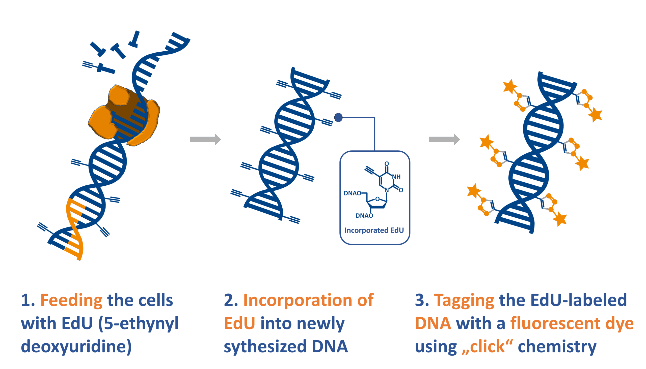
The In Vivo ClickTech EdU Cell Proliferation Kit for High-Throughput Screening (HTS) enables precise detection of cell proliferation in whole organisms, supporting studies on tumor growth, neural and tissue development, and cellular regeneration. The assay uses the thymidine analogue EdU (5-ethynyl-2′-deoxyuridine), incorporated into newly synthesized DNA during the S-phase of the cell cycle. Detection is achieved through bioorthogonal ‘click’ chemistry, which fluorescently tags EdU-labeled DNA without denaturation. This ensures bright, stable signals with minimal background, ideal for high-content analysis across diverse tissue types and organisms.

How this kit differs from standard HTS kits
Unlike standard EdU HTS kits designed for cultured cells, the in vivo kit provides increased EdU quantities for systemic administration and includes protocols optimized for animal and tissue samples. Detection reagents are tailored for complex biological matrices, ensuring reproducibility and compatibility with imaging workflows.
How does the ClickTech EdU Cell Proliferation Assay work?
5-ethynyl-2′-deoxyuridine (5-EdU) is a cell-permeable derivative of thymidine in which the methyl group has been replaced by an ethynyl group. EdU is converted to the triphosphate 5-EdUTP and incorporated into newly synthesized DNA during replication. Bioorthogonal copper-catalyzed azide–alkyne cycloaddition (CuAAC) ‘click’ chemistry [1, 2] is then used to covalently link azide-modified fluorophores to the EdU for detection. This produces highly specific nuclear labeling of proliferating cells with excellent spatial resolution.[3]
Why choose ClickTech In Vivo HTS?
- Exceptional sensitivity with low background
Click chemistry-based dye conjugation ensures a high signal-to-noise ratio; no cross-reactivity to other biomolecules present in a tissue.
- Faster, simpler workflow
No DNA denaturation or antibody steps necessary.
- No harsh treatments or specialized equipment needed
Tissue morphology and antigenicity are preserved — ideal for multiplexing with immunostaining.
What are the features & applications of the in vivo ClickTech EdU HTS assay?
- Suitable for imaging in mice, rats, zebrafish, and more
- Compatible with injection, water-based delivery or incubation
- Antibody-free, fast detection (~30 minutes)
- Works with multiplex labeling and co-staining
Which organisms can the In Vivo ClickTech EdU Cell Proliferation Assay be used on?
This assay has been used with many different types of organisms, ranging from prokaryotic to eukaryotic. It can be used with animals such as mice, rats, C. elegans and zebrafish.[4]
Literature
- Peptidotriazoles on Solid Phase: [1,2,3]-Triazoles by Regiospecific Copper(I)-Catalyzed 1,3-Dipolar Cycloadditions of Terminal Alkynes to Azides. C. W. Tornøe, C. Christensen, M. Meldal, 2002, J. Org. Chem., Vol. 67, p. 3057–3064.
- A Stepwise Huisgen Cycloaddition Process: Copper(I)-Catalyzed Regioselective “Ligation” of Azides and Terminal Alkynes. V. V. Rostovtsev, L. G. Green, V. V. Fokin, K. B. Sharpless, 2002, Angew. Chemie Int. Ed., Vol. 41, p. 2596–2599.
- A novel multicolor immunostaining method using ethynyl deoxyuridine for analysis of in situ immunoproliferative response, Y. Kitazawa, H. Ueta, T. Hünig, Y. Sawanobori, K. Matsuno, 2015, Cell Biol., Vol. 144, p. 195–208.
- Skin cells undergo asynthetic fission to expand body surfaces in zebrafish, K. Y. Chan, C.-C. Sanders Yan, H.-Y. Roan, S.-C. Hsu, T.-L. Tseng, C.-D. Hsiao, C.-P. Hsu & C.-H. Chen, 2022, Nature, Vol. 605, p. 119–125.
- An efficient protocol for in vivo labeling of proliferating epithelial cells, C. Michel et al., 2018, J. Immunol. Methods, Vol. 457, p. 82-86. https://doi.org/10.1016/j.jim.2018.03.015
- Cisplatin-induced DNA double-strand breaks promote meiotic chromosome synapsis in PRDM9-controlled mouse hybrid sterility, L. Wang et al., 2018, eLife, Vol. 7, p. e42511. https://doi.org/10.7554/eLife.42511
- FXR Regulates Intestinal Cancer Stem Cell Proliferation, T. Fu et al., 2019, Cell, Vol. 176, p. 1098-1112. https://doi.org/10.1016/j.cell.2019.01.036
FAQ
-
What type of cells can incorporate EdU?
The EdU cell proliferation assay has been applied to many different cell types and organisms from prokaryotic to eukaryotic. Cell lines such as E. coli, HeLa, HEK, MOLM are arguably among the most routine applications, but also animals, like mouse, rat, the nematode C. elegans, crickets (Gryllus bimaculatus), chicken (Gallus domesticus) and zebra fish (Danio rerio) or even plants (e.g. Arabidopsis thaliana) can be applied.
Cells that possess a pyrimidine pathway that can phosphorylate EdU to the corresponding triphosphate, which is then accepted by the host DNA polymerase for incorporation into DNA during replication. -
Can I perform EdU cell proliferation detection on living cells or tissues?
EdU is incorporated into living cells, but the detection reaction must be performed on fixed and permeabilized samples.
-
How does EdU labeling compare to BrdU or the 3H-thymidine incorporation assay?
All three methods enable to determine cell proliferation directly by incorporation of a metabolite analogue and subsequent detection. The 3H-thymidine incorporation assay is very sensitive, but the radioactive compound requires specialized equipment and dedicated lab space for handling. EdU and BrdU assays are non-radioactive alternatives with decreased risk for health and environment. Compared to the BrdU incorporation assay the EdU assay is faster, more sensitive, requires less handling time and needs no harsh DNA denaturing conditions for detection. Therefore, the EdU cell proliferation is also compatible with multiplexing and preserving tissue integrity.
-
Can I combine DAPI staining and EdU detection?
Yes, this is feasible. Please note that DAPI staining should be done after the click detection step. Alternatively, SYBR Green DNA staining can be used. But, please note that SYBR green should not be used with dyes of 488 nm wavelengths.
-
When can I safely interrupt the experiment?
It is possible to safely interrupt the protocol after the fixation step. Thereto, remove the fixation solution and wash as suggested by the user manual, then the cells can be stored in buffer at 4° C. Alternatively, the experiment can also be safely interrupted after permeabilization, again as described above.
Please note: It is important to proceed with the experiment if the click cocktail for the detection of the EdU has been prepared already. -
Is antibody staining compatible with the EdU?
Antibody staining is compatible with EdU cell proliferation detection when antibody detection is done after the click detection step. Please be aware of the dye used for EdU detection. Check also the user manual for more information.
-
How to determine the EdU incubation time?
The EdU incubation time depends on the cell type or organism, the applied EdU concentration and the experimental design. For a start it is advisable to refer to a literature protocol (which is close to your experimental setup) and to test the conditions with a low number of samples. As a general guideline we recommend to use a maximum of 10 µM final EdU in the cell culture medium for incubations. For longer incubation (> 1 day) the concentration should be decreased to 1-5 µM.
-
Is this kit for diagnostic or therapeutic use?
No. This kit is for Research Use Only (RUO) and must not be used for diagnostic or clinical purposes.
-
How does the in vivo kit differ from the standard EdU HTS kit?
The in vivo kit includes higher EdU concentration for systemic delivery and protocols tailored for tissue and whole-organism samples.
-
Which organisms are compatible?
Validated for mice, rats, zebrafish, C. elegans, and other research models. Broad compatibility ensures adaptability across experimental setups.
-
Is the workflow automation-friendly?
Yes. The protocol is optimized for high-content imaging platforms, robotic liquid handling systems, and LIMS integration.
-
What safety considerations apply?
Follow standard laboratory safety protocols for handling chemicals and animal samples. The kit does not contain radioactive materials. For more information, please check our safety data sheet section