

In Vivo EdU Cell Proliferation Assay for Flow Cytometry
ClickTech In Vivo EdU Cell Proliferation Kit for Flow Cytometry
| Size | Catalog No. | Price |
|---|---|---|
| Dye 488 / 50 Assays / Size S | BCK-EdU488FC50+IV-S | € 530,00 |
| Dye 488 / 50 Assays / Size M | BCK-EdU488FC50+IV-M | € 665,00 |
| Dye 488 / 50 Assays / Size L | BCK-EdU488FC50+IV-L | € 810,00 |
| Dye 555 / 50 Assays / Size S | BCK-EdU555FC50+IV-S | € 530,00 |
| Dye 555 / 50 Assays / Size M | BCK-EdU555FC50+IV-M | € 665,00 |
| Dye 555 / 50 Assays / Size L | BCK-EdU555FC50+IV-L | € 810,00 |
| Dye 594 / 50 Assays / Size S | BCK-EdU594FC50+IV-S | € 530,00 |
| Dye 594 / 50 Assays / Size M | BCK-EdU594FC50+IV-M | € 665,00 |
| Dye 594 / 50 Assays / Size L | BCK-EdU594FC50+IV-L | € 810,00 |
| Dye 647 / 50 Assays / Size S | BCK-EdU647FC50+IV-S | € 530,00 |
| Dye 647 / 50 Assays / Size M | BCK-EdU647FC50+IV-M | € 665,00 |
| Dye 647 / 50 Assays / Size L | BCK-EdU647FC50+IV-L | € 810,00 |
Chemical Properties
-
Shelf Life
12 months unopened after receipt
-
Storage Conditions
2-8 °C
-
Physical State
kit system made of different components
-
CAS Number
n.a.
-
Excitation (max)
Dye 488: 496 nm | Dye 555: 546 nm | Dye 594: 584 nm | Dye 647: 643 nm
-
Emission (max)
Dye 488: 516 nm | Dye 555: 579 nm | Dye 594: 603 nm | Dye 647: 662 nm
-
Ɛ (max)
Dye 488: 83.000 cm-1M-1 | Dye 555: 91.000 cm-1M-1 | Dye 594: 110.000 cm-1M-1 | Dye 647: 250.000 cm-1M-1
-
Preparation/Handling
please see user manual of the kit
Product Information
Reliable, Non-Radioactive Detection of DNA Synthesis in Living Organisms
The In Vivo EdU Cell Proliferation Assay for Flow Cytometry provides a robust and non-radioactive method for tracking de novo DNA synthesis in living organisms. Unlike traditional BrdU or ³H-thymidine assays, this approach avoids harsh DNA denaturation and radioactive hazards, enabling fast, precise, and multiplex-compatible detection of proliferating cells in complex biological systems.
Principle of the Assay
EdU (5-ethynyl-2′-deoxyuridine), a thymidine analog, is administered to the organism via injection or oral dosing. During the S-phase, EdU is incorporated into replicating DNA. Detection of the incorporated EdU is achieved through a copper-catalyzed azide-alkyne cycloaddition (click chemistry), which conjugates EdU to a fluorescent azide dye without requiring DNA denaturation or antibodies. This mild and highly specific reaction preserves sample integrity and supports downstream multiplexing.
How it works:
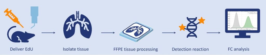
- In vivo EdU Incorporation
EdU can be delivered to animals (e.g., mice, rats, zebrafish) via intraperitoneal, intramuscular, or subcutaneous injection, orally, or through direct incubation, depending on the model. Inside cells, EdU is phosphorylated into EdU-triphosphate and integrated by DNA polymerase, marking actively dividing cells. Recommended dosing: 10 µM for short studies (2–24 hours) or 1–5 µM for longer labeling periods.
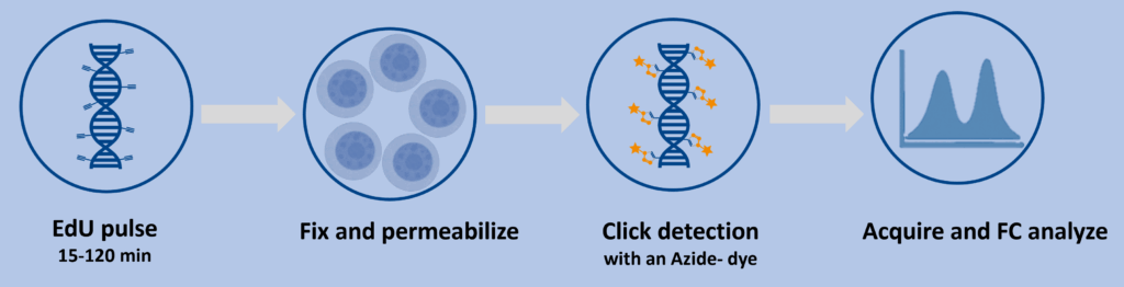
- Cell Preparation
After EdU incubation (2 hours to 1 day), harvest cells or tissues, then fix with paraformaldehyde to preserve structure and permeabilize with 0,5% saponin solution to allow reagent access to the DNA, preparing samples for detection.
- Click Chemistry Detection
A rapid click reaction conjugates EdU’s alkyne group in the DNA to a fluorescent azide dye using a copper catalyst, forming a stable triazole bond that makes dividing cells to emit a bright fluorescent signal with minimal background noise, detectable by flow cytometry.
- Flow Cytometry Analysis
Analyze labeled cells using a flow cytometer with standard channels (488, 555, 594, or 647 nm). Apply gating strategies including forward/side scatter, live/dead discrimination, and marker-based identification for accurate interpretation.
To obtain more comprehensive datasets, EdU labeling can be combined with immunohistochemistry (IHC) and DNA counterstaining. Perform EdU detection prior to immunohistochemistry (IHC) to prevent antigen loss caused by the copper-catalyzed reaction. Add DAPI or antibody staining after the click reaction to enable multiplex imaging, with DNA counterstaining typically performed last. This sequencing allows collection of richer multiparametric data while minimizing spectral overlap.
Why Choose baseclick’s In Vivo EdU Assay?
- Superior Accuracy: Direct labeling of proliferating cells without antibodies or radiation.
- Fast and Efficient: Detection completed in ~30 minutes post-fixation.
- Flexible Delivery: Injection, oral dosing, or incubation for diverse models.
- Multiplex Ready: Combine with antibody staining or nuclear dyes for comprehensive analysis.
- Broad Applicability: Suitable for mammals, fish, insects, worms, and plants.
Key Differences between In Vivo and In Vitro
The in vivo kit is designed for whole-organism studies, enabling systemic EdU uptake and tissue-specific analysis in models such as mice, zebrafish, or plants. It is ideal for drug efficacy, toxicology, and developmental biology research. In contrast, the in vitro kit is optimized for controlled cell culture experiments, supporting high-throughput screening and rapid drug testing. Both kits use the same reliable click chemistry and flow cytometry detection for consistent, high-quality results.
Advantages
The In Vivo EdU Cell Proliferation Assay offers significant advantages over traditional methods:
- Non-Radioactive and Safe: Eliminates hazards of ³H-thymidine.
- Gentle on Samples: No harsh DNA denaturation as required for BrdU.
- High Sensitivity and Specificity: Detects proliferating cells across diverse organisms.
- Rapid and Efficient: The EdU detection process takes approximately 30 minutes, streamlining workflows and saving valuable time.
- Versatile and Flexible: Available in 3 kit sizes and compatible.
- Multiplexing Capability: Compatible with immunostaining and nuclear dyes.
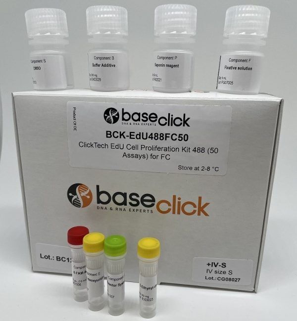
Kit Contents
- EdU (S, M, or L sizes)
- Fluorescent dye (488, 555, 594, or 647 nm)
- Click reaction reagents
- Fixation and permeabilization solutions
- Clear protocol for in vivo and flow cytometry workflows
Tips for Success
- Use 10 µM EdU for short-term labeling or 1–5 µM for extended studies.
- Store fixed samples at 4 °C for flexible analysis timing.
- Perform EdU detection before antibody staining to preserve epitopes.
LITERATURE
An efficient protocol for in vivo labeling of proliferating epithelial cells, C. Michel et al., 2018, J. Immunol. Methods, Vol. 457, p. 82-86.
https://doi.org/10.1016/j.jim.2018.03.015
Cisplatin-induced DNA double-strand breaks promote meiotic chromosome synapsis in PRDM9-controlled mouse hybrid sterility, L. Wang et al., 2018, eLife, Vol. 7, p. e42511.
https://doi.org/10.7554/eLife.42511
FXR Regulates Intestinal Cancer Stem Cell Proliferation, T. Fu et al., 2019, Cell, Vol. 176, p. 1098-1112.
https://doi.org/10.1016/j.cell.2019.01.036
A modified method to analyse cell proliferation using EdU labelling in large insect brains, A. Anton et.al., 2023, PLoS One, Vol. 18(10), p. e0292009.
PLoS One; doi: 10.1371/journal.pone.0292009. eCollection 2023.
EdU-Based Assay of Cell Proliferation and Stem Cell Quiescence in Skeletal Tissue Sections, M. Angelozzi et al., 2021, Methods in Molecular Biology, Vol. 2230, p. 357–365.
FAQ
-
How should I administer EdU in vivo?
EdU can be delivered via intraperitoneal, intramuscular, or subcutaneous injection, oral dosing, or direct incubation, depending on the organism and experimental design. For short-term labeling (30 min – 2 hours), start with 10 µM; for longer studies, use 1–5 µM.
-
Which organisms can I use this kit with?
The assay is compatible with a wide range of models, including mammals (e.g., mice, rats), zebrafish, insects, worms, and plants—any system with active DNA synthesis.
-
How long should I incubate EdU in vivo?
Incubation time depends on the proliferation rate of the target tissue. Common ranges are 30 min to 4 hours (Zebrafish embryos or larvae). For slow-dividing cells (e.g., certain stem cell populations, adult neurons in rare cases), longer exposure may be necessary.
-
Can I combine EdU detection with immunostaining or DNA counterstaining?
Yes. Perform the EdU click reaction first, then proceed with antibody staining or DAPI counterstaining. This ensures antigen integrity and avoids interference from the copper catalyst.
-
What fluorophores are available?
The kit offers fluorescent azide dyes for common flow cytometry channels:
488 nm (FITC channel), 555 nm, 594 nm, 647 nm
-
How long does the detection step take?
The click chemistry detection is rapid and typically completed in about 30 minutes after fixation and permeabilization.
-
Is the assay safe compared to ³H-thymidine?
Yes. EdU is non-radioactive, eliminating health and environmental hazards associated with radioactive labeling.
-
How should I store the kit and prepared samples?
Store the kit at 2–8 °C in the dark. Fixed samples can be stored at 4 °C for several days before analysis.