

Proliferation Spot T-Cell Kit for Imaging
Dual Detection of DNA Synthesis and Antigen-Specific Immune Function
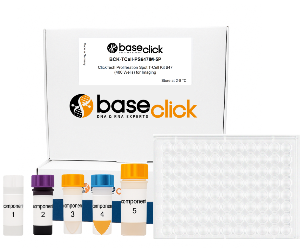
| Size | Catalog No. | Price |
|---|---|---|
| Dye 488 / 480 Wells | BCK-TCell-PS488IM-5P | € 845,00 |
| Dye 647 / 480 Wells | BCK-TCell-PS647IM-5P | € 845,00 |
Chemical Properties
-
Shelf Life
12 months unopened after receipt
-
Storage Conditions
2-8 °C
-
Physical State
kit system made of different components
-
CAS Number
n.a.
-
Excitation (max)
Dye 488: 496 nm | Dye 647: 643 nm
-
Emission (max)
Dye 488: 516 nm | Dye 647: 662 nm
-
Ɛ (max)
Dye 488: 83.000 cm-1M-1 | Dye 647: 250.000 cm-1M-1
-
Preparation/Handling
please see user manual of the kit
Product Information
Introduction
The ClickTech Proliferation Spot T-Cell Kit (ProliSpot) is an innovative assay platform that combines two complementary principles in one workflow:
- EdU-based DNA synthesis detection for precise identification of proliferating T cells.
- ELISpot-based cytokine capturing for antigen-specific functional profiling of immune cells at a single cell level.
ProliSpot is an integrated approach that enables simultaneous measurement of cell division and immune activation at single-cell resolution without radioactive labeling, harsh DNA denaturation, or the need for flow cytometry analysis.
Kit Variants
- ClickTech Proliferation Spot T-Cell Kit 488 (480 Wells) for Imaging
FITC channel (~488 nm) for fluorescence detection
- ClickTech Proliferation Spot T-Cell Kit 647 (480 Wells) for Imaging
Cy5 channel (~647 nm) for far-red detection
Both kits are optimized for automated imaging, high-throughput analysis, and single-cell resolution, making them suitable for:
- Immunological research
- Vaccine development
- CAR-T potency testing
- Autoimmunity and cancer immunology studies
Scientific Background
EdU Assays: Detect DNA replication by incorporating 5-ethynyl-2′-deoxyuridine (EdU) into newly synthesized DNA, visualized via Click Chemistry. Advantages include high sensitivity and no need for harsh DNA denaturation (unlike necessary in BrdU based assays).
ELISpot Assays: Quantify cytokine secretion by capturing proteins on antibody-coated membranes, visualized as discrete spots. ELISpot is highly sensitive for immune function but does not measure proliferation.
ProliSpot combines both, enabling researchers to correlate proliferation and antigen-specific cytokine secretion in the same well.
Materials and Methods
- Cell Preparation: Peripheral blood lymphocytes (PBLs) were stimulated for 48 hours with S. aureus Toxin E at 250,000 cells per well.
- Plate Setup: Cells were coated onto ProliSpot-prepared glass-bottom plates with PVDF membranes and PEI activation for optimal protein binding.
- EdU Detection: Incorporation of EdU was visualized via copper(I)-catalyzed azide-alkyne cycloaddition (Click Chemistry) using 6-FAM Picoly azide and Eterneon Red Picoly azide.
- Imaging: Automated imaging performed with a 10× air objective, 6×6 mosaic acquisition, and stitching.
- Analysis: Image segmentation, cell counting, and fluorescence intensity measurement (EdU-488 per cell) were conducted using automated software.
Results
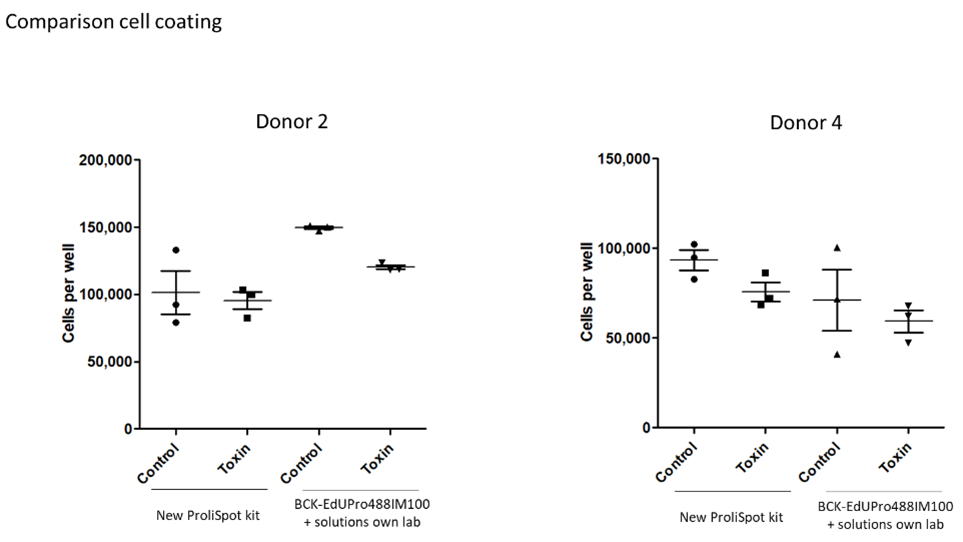
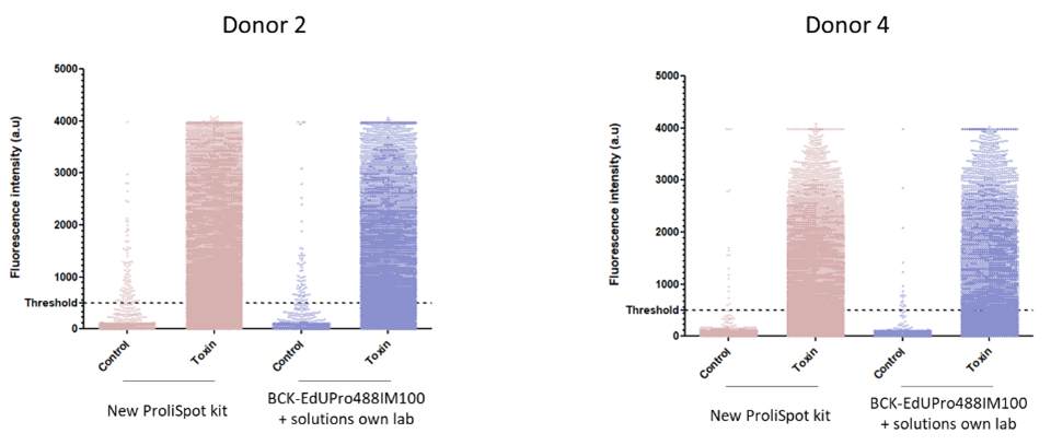
ClickTech Proliferation Spot T-Cell Kit (ProliSpot) for ELISpot analysis was developed using baseclicks Sensitive EdU Cell Proliferation Kit for Imaging (BCK-EdUPro488IM100). The protocol and solutions were in particular adapted on plate coating efficiency and subsequent fluorescent detection quality. After establishing the procedure for the ELISpot detection and manufacturing process we compared the performance.
Coating Efficiency: Comparison between the manufactured ProliSpot kit and the adapted procedure for ELISpot detection basis of BCK-EdUPro488IM100 on adhesion efficiency (Figure 1) and detection capacity (Figure 2) showed almost identical performance of the kit to the established lab procedure.
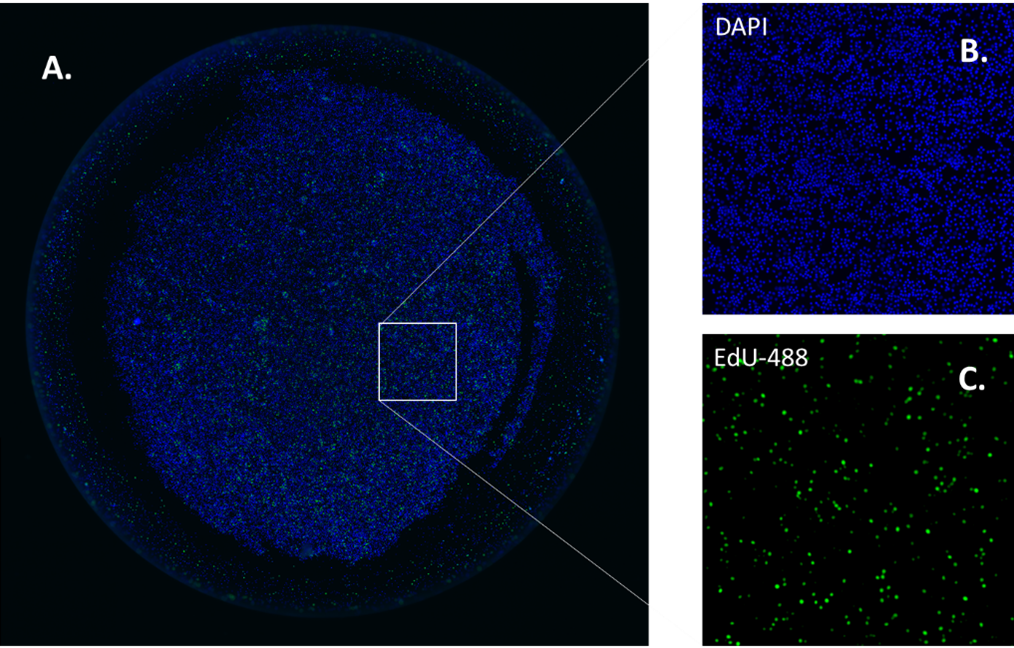
Figure 1: Images of cell adhesion on coated plates. Generally 100.000 cells per well were analyzed and constant adhesion performance was observed from different blood donors for the ClickTech Proliferation Spot T-Cell Kit (ProliSpot) and the laboratory procedure. A. Image of a well DAPI emission (blue cell nucleus). B. Close up DAPI emission (blue cell nucleus) C. Close up FAM emission (green cells).
A.
B.
C.

Figure 2: baseclick’s newly developed ClickTech Proliferation Spot T-Cell Kit (ProliSpot) exhibited similar performance in A. adhesion of cells to the plate surface, B. fluorescence intensity per well and C. toxin stimulation evaluation testing.
Discussion
ProliSpot demonstrated high performance in cell coating, EdU incorporation and fluorescence detection allowing automated imaging capturing in ELISpot machines.
Conclusion
The ClickTech Proliferation Spot T-Cell Kit offers a sensitive, reliable, and scalable method for T-cell proliferation analysis. Its dual-readout capability, DNA synthesis and antigen-specific cytokine detection, provides a comprehensive view of immune responses, making it a powerful tool for advanced immunological research, diagnostic and drug development.
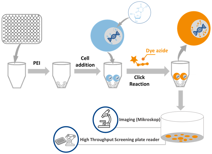
Workflow Overview
- Cell stimulation
- EdU incorporation
- ProliSpot plate preparation
- Cell transfer & fixation
- Click Chemistry detection (FITC or Cy5)
- Automated imaging & analysis
Technical Summary
| Parameter | Specification |
| Detection Principles | EdU incorporation (Click Chemistry) + cytokine capture |
| Format | 96-well PVDF plates |
| Fluorescent Channels | baseclick dyes: FITC (green, ~488 nm) or Cy5 (far-red, ~647 nm |
| Cell Input | 250,000 cells per well (optimized for PBMC/PBL) |
| Workflow Compatibility | Automated imaging, mosaic stitching, segmentation |
| Readouts | % EdU-positive cells + SFU count per antigen |
| Sensitivity | Detects rare antigen-specific T cells (1 in 10⁶) |
Comparison of Methods
| Feature | Standard EdU Kit (baseclick) | ELISpot | ProliSpot Kit |
| Detection | DNA synthesis (EdU) | Cytokine secretion | DNA synthesis + cytokine secretion |
| Format | Suspension/monolayer | Antibody-coated membrane | Glass-bottom plate, spot-based |
| Readout | Flow cytometry/microscopy | Spot counting | Imaging + SFU |
| Application | General proliferation | Immune function (cytokines) | T-cell proliferation, high-throughput |
Kit Components
- EdU reagent
- Fluorescent azide dye (488 or 647)
- DMSO
- Reaction Buffer
- Reactor system
- Buffer Additive
- Saponin
- BSA-c
- ProliSpot PVDF plates
- Polyethylenimine (PEI) coating solution
- DAPI nuclear stain
- Glutaraldehyde
- Detailed protocol for human T-cell workflows
Key Advantages
- Dual Readout: Proliferation + immune function in one assay
- High Sensitivity: Detects rare antigen-specific T cells
- Automation Ready: Compatible with high-throughput imaging systems
- Safe & Efficient: No radioactive labeling or harsh DNA denaturation
FAQ
-
What makes ProliSpot different from standard EdU kits?
ProliSpot combines EdU-based DNA synthesis detection with spot-based cytokine analysis, enabling simultaneous measurement of T-cell proliferation and antigen-specific immune function in the same well. Standard EdU kits only measure proliferation and require separate assays for functional analysis.
-
How does ProliSpot compare to ELISpot?
ELISpot detects cytokine secretion but does not measure cell proliferation. ProliSpot integrates both, providing a more comprehensive view of immune responses without additional assays.
-
What fluorescent dyes are included?
The kit uses baseclick fluorescent dyes: FITC (green, ~488 nm) and Cy5 (far-red, ~647 nm)
Choose the variant based on your imaging system and multiplexing needs.
-
Is the kit compatible with automated imaging systems?
Yes. ProliSpot is optimized for high-throughput imaging workflows, including mosaic stitching and automated segmentation.
-
What is the recommended cell input per well?
The protocol is optimized for 250,000 PBMCs or PBLs per well, but adjustments can be made based on experimental design.
-
Does the assay require DNA denaturation or radioactive labeling?
No. ProliSpot uses Click Chemistry, which is safe, non-radioactive, and does not require harsh DNA denaturation steps.
-
Can ProliSpot detect rare antigen-specific T cells?
Yes. The assay can detect rare proliferating T cells at frequencies as low as 1 in 10⁶, making it suitable for vaccine and immunotherapy studies.
-
What applications is ProliSpot best suited for?
Vaccine development
CAR-T potency testing
Autoimmunity and cancer immunology
Drug screening for immunomodulatory compounds