


ClickTech Proliferation Spot T-Cell Kit
High-Sensitivity Detection of Rare Cell Proliferation Events
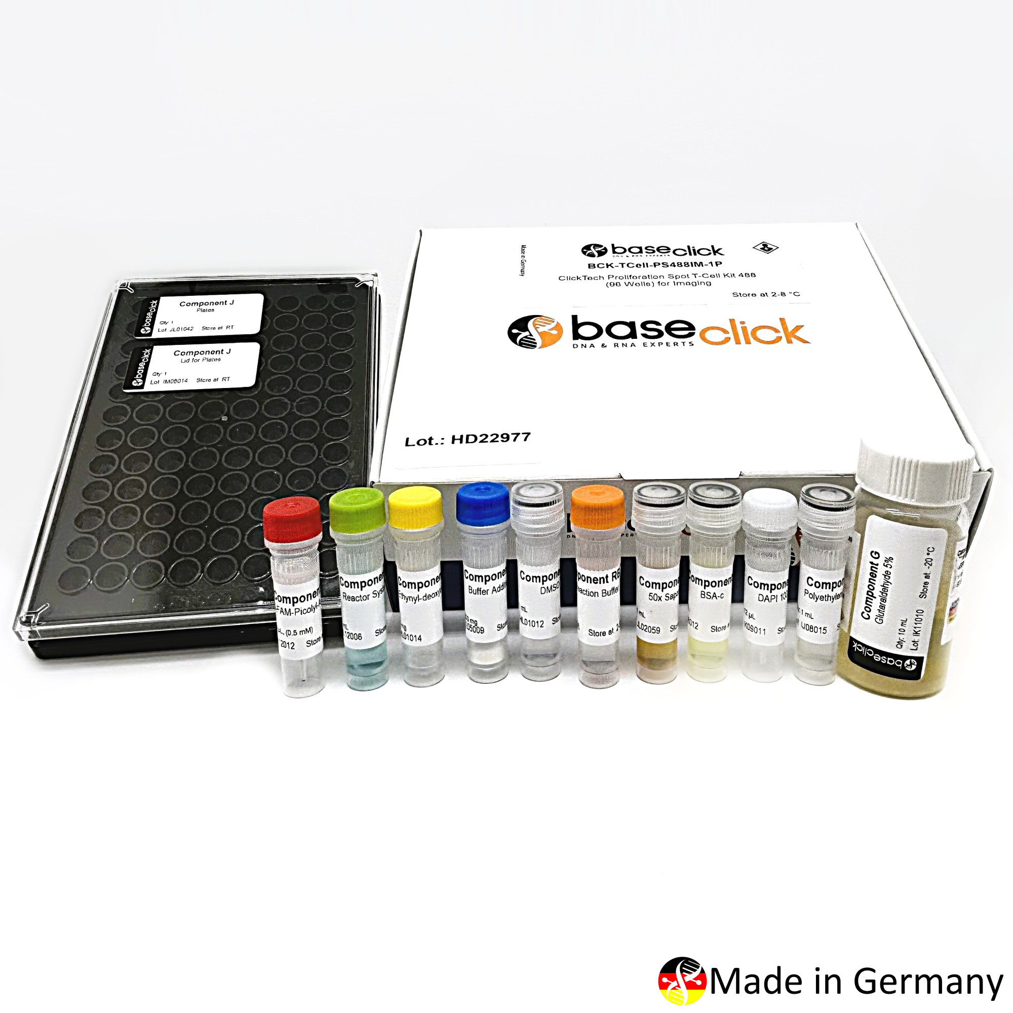
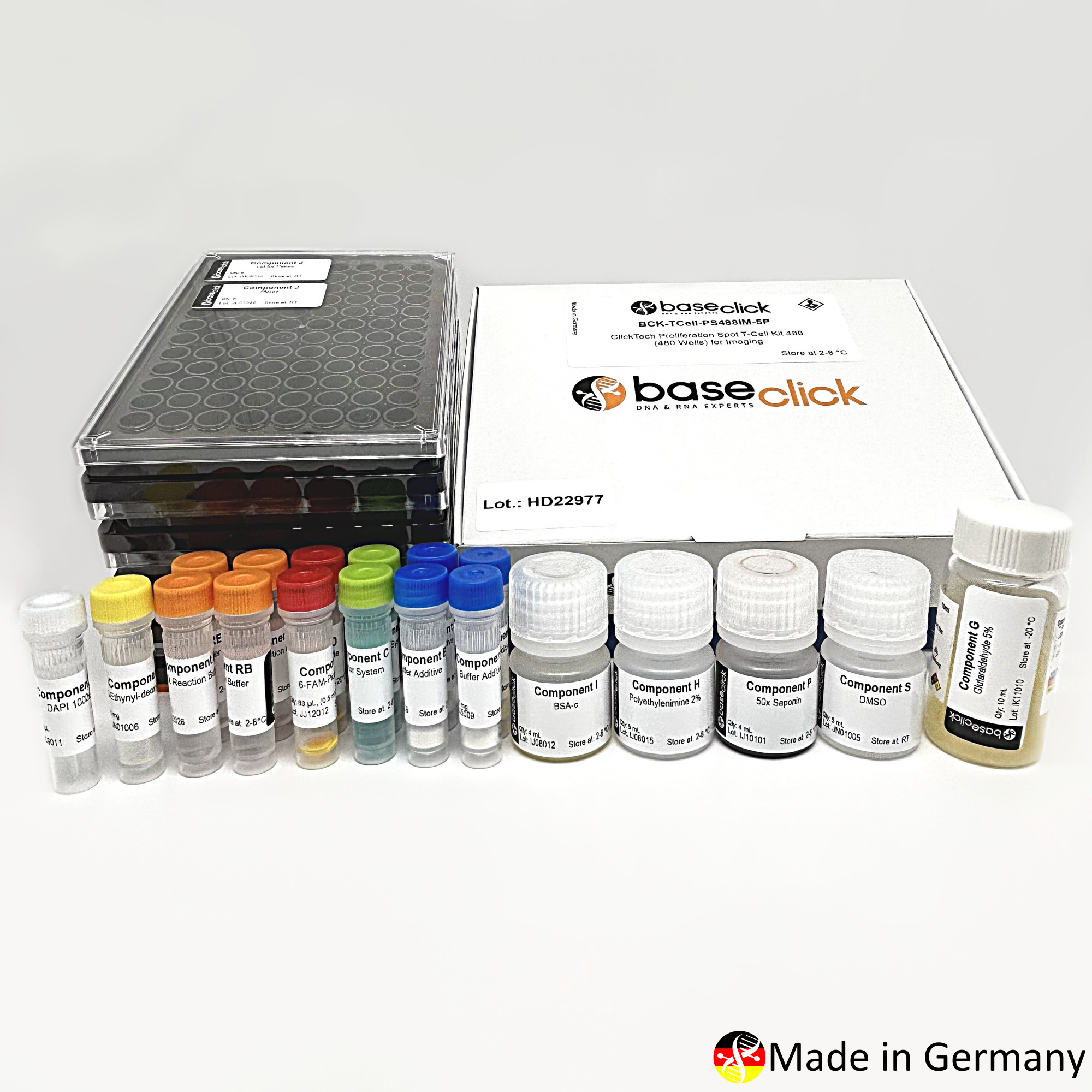


| Size | Catalog No. | Price |
|---|---|---|
| Dye 488 / 96 Wells | BCK-TCell-PS488IM-1P | € 320,00 |
| Dye 488 / 480 Wells | BCK-TCell-PS488IM-5P | € 845,00 |
| Dye 647 / 96 Wells | BCK-TCell-PS647IM-1P | € 320,00 |
| Dye 647 / 480 Wells | BCK-TCell-PS647IM-5P | € 845,00 |
Chemical Properties
-
Shelf Life
12 months unopened after receipt
-
Storage Conditions
2-8 °C
-
Physical State
kit system made of different components
-
CAS Number
n.a.
-
Excitation (max)
Dye 488: 496 nm | Dye 647: 643 nm
-
Emission (max)
Dye 488: 516 nm | Dye 647: 662 nm
-
Ɛ (max)
Dye 488: 83.000 cm-1M-1 | Dye 647: 250.000 cm-1M-1
-
Preparation/Handling
please see user manual of the kit
Product Information
Introduction
The ClickTech Proliferation Spot T-Cell Kit (ProliSpot) is an innovative assay platform that combines two complementary technologies in one workflow:
- EdU-based DNA synthesis detection for precise identification of rare proliferating cells e.g. T cells.
- Covalent cell immobilization on a solid support for highly sensitive detection and enumeration of rare proliferating antigen-responsive cells.
ProliSpot is an integrated approach that enables measurement of cell proliferation and can also be multiplexed with activation markers such as CD137 or CD154, without radioactive labeling, harsh DNA denaturation, or the need for flow cytometry analysis.
Kit Variants
- ClickTech Proliferation Spot T-Cell Kit 488 (480/96 Wells) for Imaging
FITC channel (~488 nm) for fluorescence detection
- ClickTech Proliferation Spot T-Cell Kit 647 (480/96 Wells) for Imaging
Cy5 channel (~647 nm) for far-red detection
Both kits are optimized for automated imaging, high-throughput analysis, and single-cell resolution, making them suitable for:
- Rare cell division experiments e.g. in blood cancer studies
- Immunological research
- Vaccine development
- CAR-T potency testing
- Autoimmunity and cancer immunology studies
Scientific Background
EdU Assays: Detect DNA replication by incorporating 5-ethynyl-2′-deoxyuridine (EdU) into newly synthesized DNA, visualized via Click Chemistry. Advantages include high sensitivity and no need for harsh DNA denaturation (unlike necessary in BrdU based assays).
Example: Materials and Methods
- Cell Stimulation and EdU Labeling: Peripheral blood mononuclear cells (PBMCs) or peripheral blood lymphocytes (PBLs) were cultured in 96-well plates and stimulated with the appropriate antigen for 24 h. A 1× EdU working solution was added during the final 2 h of incubation to label proliferating cells through incorporation into newly synthesized DNA.
- ProliSpot Plate Preparation and Cell Immobilization: Glass-bottom ProliSpot plates were coated with polyethyleneimine (PEI) and subsequently activated with glutaraldehyde. Stimulated cells were transferred to the activated plates and immobilized on the glass surface by centrifugation, enabling high-density cell attachment and minimizing cell loss during subsequent washing and staining procedures.
- Cell Fixation, Permeabilization, and EdU Detection: Immobilized cells were fixed with formaldehyde in PBS, permeabilized using a saponin-containing buffer, and subjected to copper(I)-catalyzed azide-alkyne cycloaddition (Click Chemistry). Incorporated EdU was detected with either 6-FAM Picoly azide or Eterneon Red Picolyl azide.
- Counterstaining and Imaging: Cell nuclei were counterstained with DAPI. Fluorescence imaging was performed using an automated widefield or confocal microscope equipped with autofocus and motorized stage capabilities, allowing analysis of entire 96-well plates.
- Analysis: Total cells were quantified based on DAPI staining (DAPI-positive), while proliferating cells were identified by EdU-specific fluorescence. Automated image segmentation and quantification were used to determine the frequency and distribution of antigen-responsive proliferating T cells.
Results
The ClickTech Proliferation Spot T-Cell Kit (ProliSpot) was developed to enable highly sensitive detection of rare cell division in an ELISpot-like imaging format. The assay combines EdU incorporation with covalent cell immobilization on high-density glass-bottom plates, followed by fluorescence-based detection using Click Chemistry.
Protocol parameters, plate-coating conditions, and detection reagents were optimized to maximize cell retention, reduce processing-related cell loss, and improve assay sensitivity and fluorescent signal quality. Following establishment of the immobilization and imaging workflow, assay performance was evaluated by assessing the detection and quantification of rare proliferating T-cell populations. The ProliSpot approach enabled sensitive measurement of antigen-induced proliferation while supporting multiparametric analysis through optional fluorescent labeling of additional cellular markers. Compared with conventional flow cytometry-based proliferation assays, the ProliSpot format provided enhanced sensitivity for detecting low-frequency proliferative responses and allowed high-content imaging-based analysis of individual cells.
Coating Efficiency: Comparison between the manufactured ProliSpot kit and the adapted procedure for ProliSpot detection basis of BCK-EdUPro488IM100 on adhesion efficiency (Figure 1) and detection capacity (Figure 2) showed almost identical performance of the kit to the established lab procedure.
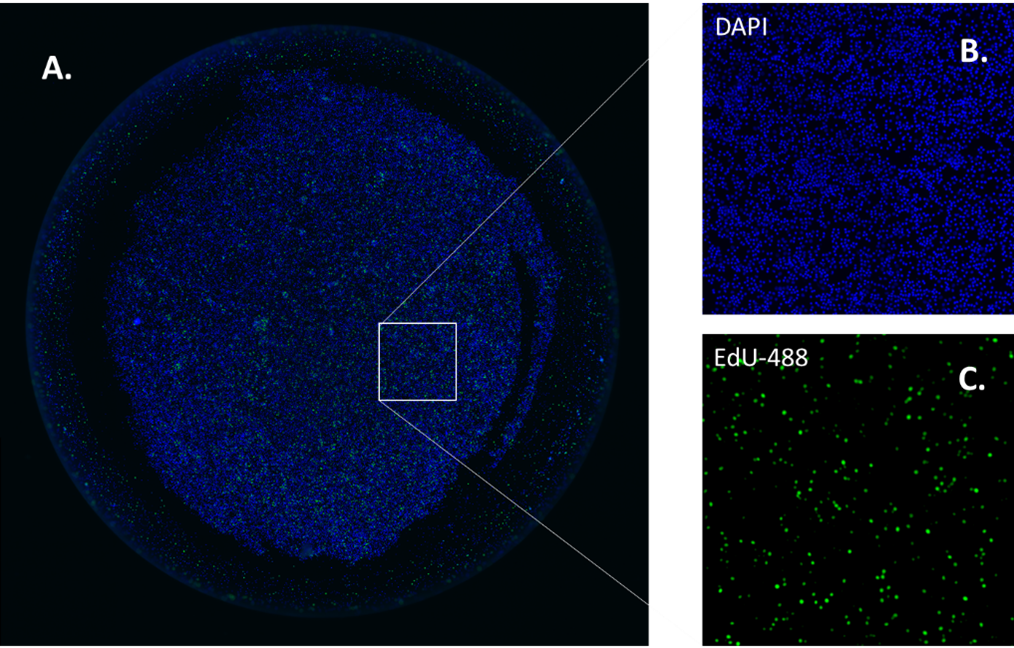
Figure 1: Images of cell adhesion on coated plates. Generally, 100,000 cells per well were analyzed and constant adhesion performance was observed from different blood donors for the ClickTech Proliferation Spot T-Cell Kit (ProliSpot) and the laboratory procedure. A. Image of a well DAPI emission (blue cell nucleus). B. Close up DAPI emission (blue cell nucleus) C. Close up FAM emission (green cells).
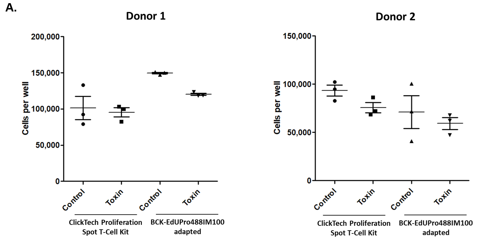
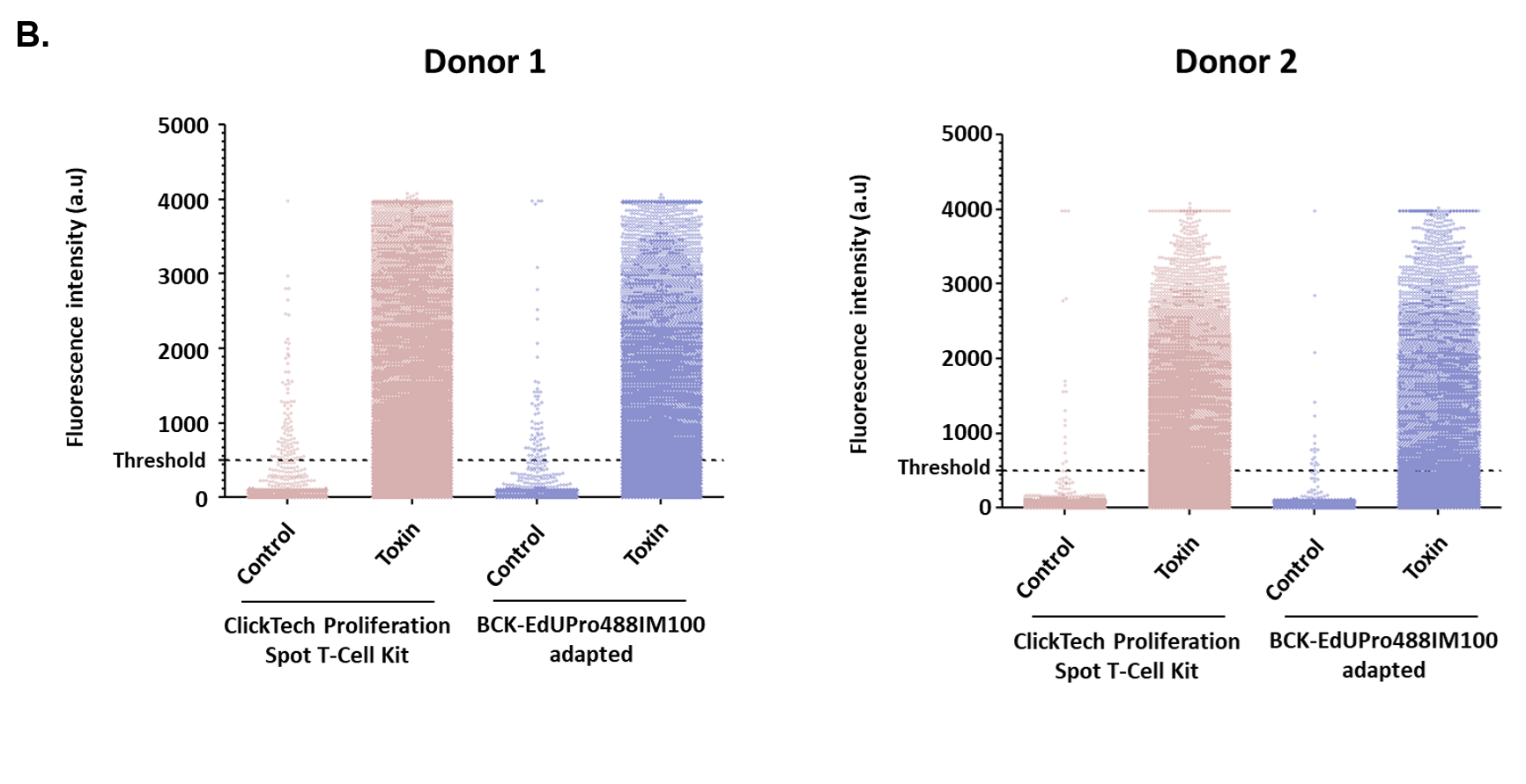
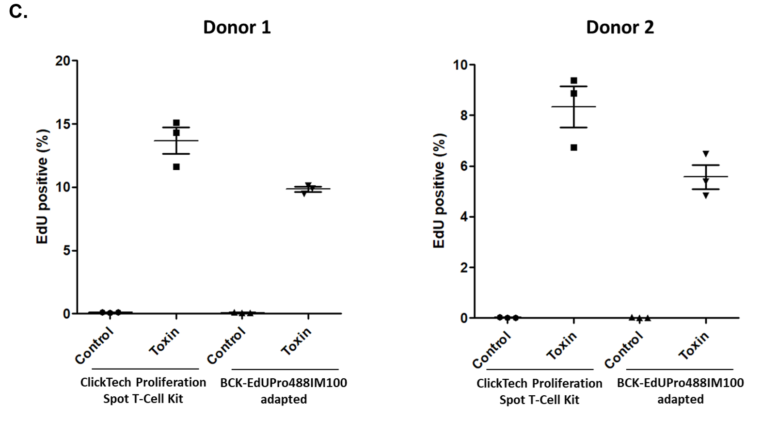
Figure 2: baseclick’s newly developed ClickTech EdU T Cell Proliferation Kit (ProliSpot) exhibited similar performance in A. adhesion of cells to the plate surface, B. fluorescence intensity per well and C. toxin stimulation evaluation testing.
Discussion
ProliSpot demonstrated robust cell immobilization, efficient EdU incorporation, and sensitive fluorescence detection, enabling reliable identification of rare proliferating cells. The assay supports multiparametric immune profiling and is compatible with fluorescence microscopy, high-content imaging systems, and FluoroSpot-based readout platforms.
Conclusion
The ClickTech Proliferation Spot T-Cell Kit offers a sensitive, reliable, and scalable platform for the detection of proliferating T cells. By combining EdU-based proliferation analysis with covalent cell immobilization and fluorescence-based detection, ProliSpot enables the sensitive quantification of rare proliferative responses and supports multiparametric immune profiling across a wide range of research applications.
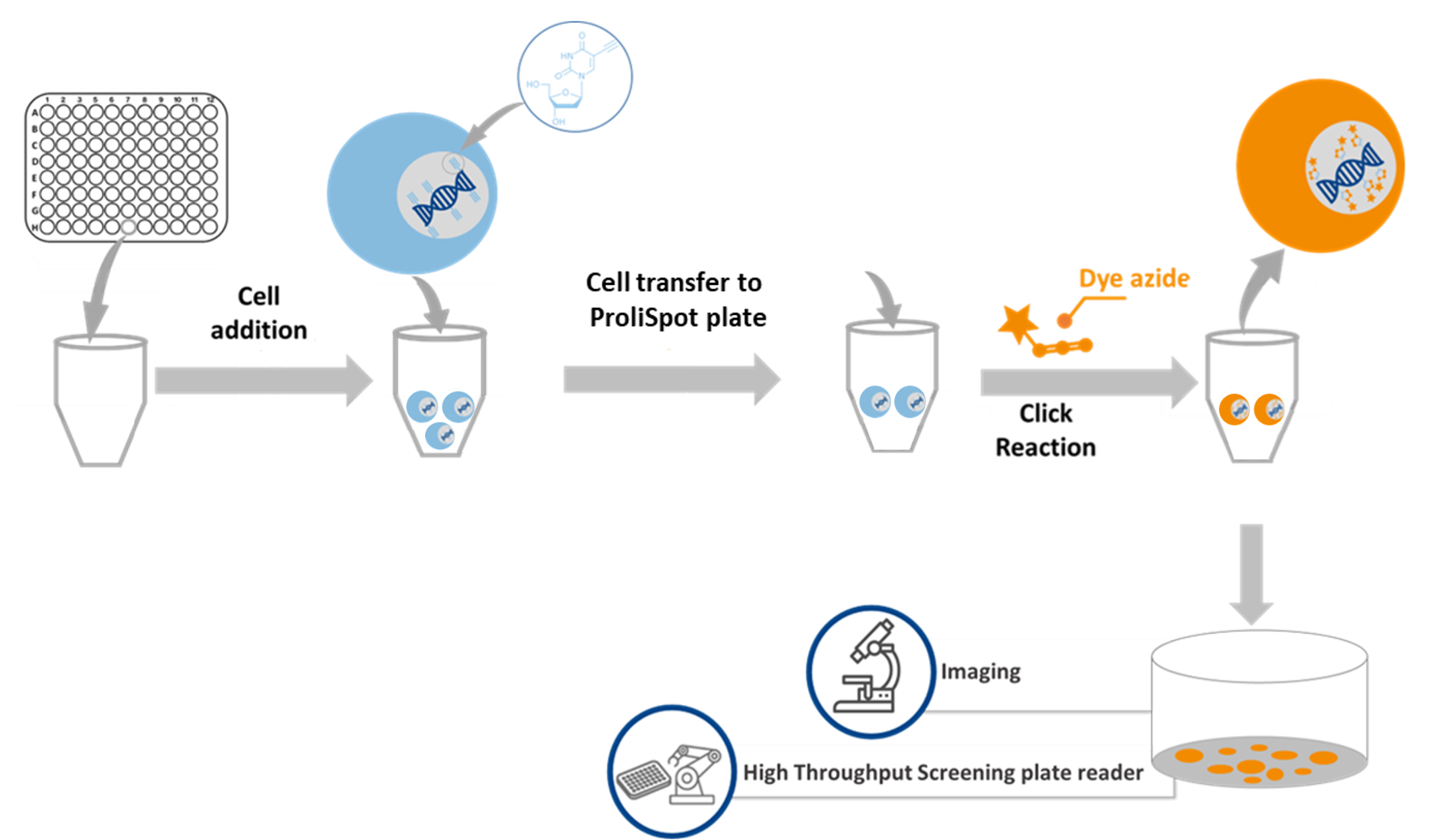
Workflow Overview
- Cell isolation
- Cell stimulation if needed
- EdU incorporation
- ProliSpot plate preparation
- Cell transfer & fixation
- Click Chemistry detection (FITC or Cy5)
- Automated imaging & analysis
Technical Summary
| Parameter | Specification |
| Detection Principles | EdU incorporation + click chemistry detection following covalent cell immobilization |
| Format | 96-well black/clear flat-bottom plates |
| Detection Dyes | 6-FAM Picolyl-Azide (Ex 496 nm / Em 516 nm) or Eterneon-Red 645 Picolyl-Azide (Ex 643 nm / Em 662 nm) |
| Cell Input | Cell inputs ranging from 100,000 to 1.5 × 10⁶ PBLs per well |
| Workflow Compatibility | Widefield microscopy, confocal microscopy, automated imaging systems, high-content screening platforms |
| Readouts | Total cell count (DAPI), EdU-positive cell count, percentage of proliferating cells, multiparametric marker co-expression |
| Sensitivity | Detects rare antigen-specific T cells division with sensitivity around 0.01%. |
Comparison of Methods combining baseclick EdU proliferation assay with ELISpot detection method
| Feature | Standard EdU Kit (baseclick) | ELISpot | ProliSpot Kit |
| Detection | DNA synthesis (EdU) | Cytokine secretion | Proliferation detection via EdU incorporation |
| Format | Suspension/monolayer | Antibody-coated membrane | Glass-bottom plate, spot-based |
| Readout | Flow cytometry/microscopy | Spot counting | Imaging + SFU |
| Application | General proliferation | Immune function (cytokines) | T-cell proliferation, high-throughput |
Kit Components
- EdU reagent
- Fluorescent azide dye (488 or 647)
- DMSO
- Reaction Buffer
- Reactor system
- Buffer Additive
- Saponin
- BSA-c
- 96-well plates
- Polyethylenimine (PEI) coating solution
- DAPI nuclear stain
- Glutaraldehyde
- Detailed protocol for human T-cell workflows
Key Advantages
- High-sensitivity detection of rare proliferating cells
- Simultaneous analysis of cell proliferation and activation markers
- No antibody-coated capture surfaces required
- Robust, reproducible, and compatible with multiple detection platforms
- Compatible with microscopy, high-content imaging, and FluoroSpot-based systems
- No radioactive labeling or harsh DNA denaturation
FAQ
-
What makes ProliSpot different from standard EdU kits?
ProliSpot combines EdU incorporation with covalent immobilization of cells at high density, enabling highly sensitive detection of proliferating cells.
-
How does ProliSpot compare to ELISpot?
ELISpot measures cytokine secretion whereas ProliSpot measures cellular proliferation. The two methods provide complementary information about antigen-specific immune responses.
-
What fluorescent dyes are included?
The kit uses baseclick’s fluorescent dyes: FITC (green, ~488 nm) and Cy5 (far-red, ~647 nm)
Choose the variant based on your imaging system and multiplexing needs.
-
Is the kit compatible with automated imaging systems?
Yes. ProliSpot is optimized for high-throughput imaging workflows, including mosaic stitching and automated segmentation.
-
What is the recommended cell input per well?
The optimal cell number depends on the application. Cell inputs ranging from 100,000 to 1.5 × 10⁶ PBLs per well have been successfully used. The appropriate cell number should be determined based on the sample type and experimental design.
-
Does the assay require DNA denaturation or radioactive labeling?
No. ProliSpot uses Click Chemistry, which is safe, non-radioactive, and does not require harsh DNA denaturation steps.
-
Can ProliSpot detect rare antigen-specific T cells?
ProliSpot has demonstrated sensitivity for detecting rare proliferating T cells at frequencies of approximately 0.01%.
-
What applications is ProliSpot best suited for?
Rare cell division e.g. in fully differentiate tissue, blood cancer diseases, Vaccine and infectious disease research, including latent tuberculosis infection (LTBI).
Cancer immunotherapy, CAR-T cell therapy monitoring, immune monitoring, and minimal residual disease (MRD).
Autoimmune and type IV hypersensitivity responses, including autoreactive T cells in Neurodegenerative disease research.