Kit for cell proliferation assay: Applications, techniques & advantages
Cell proliferation assay kits have been developed to measure cell division, viability and cytotoxicity in drug screening, cancer research and growth factor studies. These kits assess metabolic activity and DNA synthesis, providing quantitative data on cell proliferation and other markers of actively dividing cells.
Key Features
– Non-Radioactive: Safer alternatives to [3H]-thymidine (e.g., MTT, CCK-8, BrdU, EdU).
– Sensitive: Detect low cell numbers (25–50 cells).
– Simple: Minimization of steps.
– Versatile: usage in various cell types; colorimetric, fluorescent, or luminescent readouts.
– Multiplexing: Some of the kits for cell proliferation assays (e.g., CCK-8, EdU) enable complex assays to be performed on the same cells.
Common Components
– Detection Reagents: Tetrazolium salts (MTT, MTS, WST-1, CCK-8), BrdU/EdU, or fluorescent dyes (CyQUANT, alamarBlue).
– Solubilization Solutions: For example DMSO for MTT formazan.
– Antibodies: For BrdU detection.
– Buffers/Standards: For stability and calibration.
– Controls: Validate assay performance.
Working Principles:
- Metabolic (e.g., MTT, CCK-8): Viable cells convert reagents to detectable signals.
- DNA Synthesis (BrdU, EdU): Detect DNA-incorporated analogs.
- ATP-Based (CellTiter-Glo): ATP drives luminescence.
- Dye-Based (CyQUANT, CFSE): DNA binding or division tracking via fluorescence.
Introduction to kits for cell proliferation assay
Standardized cell proliferation assays are essential in modern life sciences because they:
- Ensure reproducibility across labs and experiments.
- Enable accurate drug screening and toxicity testing.
- Support cancer and regenerative research by reliably tracking cell growth.
- Facilitate high-throughput analysis, saving time and resources.
- Provide quantitative data for regulatory and clinical decisions.
- Reduce variability to minimize experimental errors and inconsistencies.
Core principles of kits for cell proliferation assay
Biological markers (biomarkers) are specific biological indicators used to assess cell proliferation and other cellular processes. Key biomarkers for cell proliferation include:
- DNA Synthesis: It detects actively dividing cells by incorporating nucleotide analogs, such as EdU, into newly synthesized DNA, which are then visualized through labeling or imaging—marking cells in the S-phase of the cell cycle.
- Learn more about EdU Assay Kit here: EdU Assay Kit Glossary
- Metabolic Activity: Active cell growth often correlates with high metabolic activity. Cell viability and proliferation can be assessed via evaluation of metabolic processes such as ATP production or reduction of tetrazolium salts (e.g., MTT, XTT).
- Cell Cycle Proteins such as proliferating cell nuclear antigen (e.g., Ki-67 or PCNA) are used to detect actively cycling cells via immunostaining or flow cytometry.
- Enzyme Activity reflects cell health and proliferation and often measured in cytotoxicity or viability assays. A commonly used enzyme activity-based assay is the lactate dehydrogenase (LDH) activity assay.
- Mitochondrial Function: Assays such as resazurin reduction (e.g. Alamar Blue) measure mitochondrial activity, which is linked to cell proliferation and energy metabolism.
These biomarkers enable quantitative, reproducible insights into cell growth, supporting research in cancer, drug development, and regenerative medicine.
Role of Biological Markers in Cell Health, Growth, and Division:
– Cell Health: Assessing biomarkers plays a pivotal role in evaluating cell viability, stress and apoptosis. For example, ATP levels and LDH release are used to assess cell viability, while caspase activity and Annexin V are used to detect apoptosis. ROS and/or glutathione are used to identify stress.
– Growth Rates: DNA synthesis assays (BrdU or EdU) and metabolic activity analyses (such as MTT or Alamar Blue) can be used to track cell population expansion. These assays are used to quantify proliferation in cancer, toxicology and regenerative studies.
– Division Cycles: Cyclins, phospho-histone H3, Ki-67, PCNA, DNA content (propidium iodide) are used to monitor cell cycle phases (G0/G1, S, G2 and M) in studies of cell cycle regulation or the effects of inhibitors.
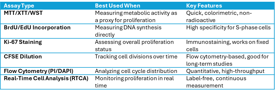
Choosing the Right Assay for Cell Proliferation

Applications of kits for cell proliferation assay in high throughput screening
Cell proliferation assay kits are widely used in high-throughput screening (HTS) to evaluate cell growth and viability, particularly in drug discovery. Based on metabolic activity or DNA synthesis, these assays enable the rapid, automated assessment of thousands of compounds. In the early stages of drug discovery, cell proliferation assays are vital for evaluating the efficacy and cytotoxicity of compounds. These assays help to identify compounds that promote or inhibit cell growth, which is essential for research in areas such as cancer, regenerative medicine and antimicrobials.
Applications of Assay Kits in HTS:
- Drug Discovery & Development
- Cancer Research
- Toxicology Testing
- Regenerative Medicine
- Personalized Medicine
Integration in Automated HTS Labs
Commercially available assay kits, such as MTT, BrdU, EdU, ATP-based assays, are optimized for automation and miniaturization. They are compatible with robotic liquid handlers and multiwell plate readers, enabling:
- Rapid screening of thousands of compounds
- Standardized, reproducible results
- Real-time or endpoint analysis with minimal manual intervention
These kits streamline the screening process, reduce variability, and accelerate the identification of promising therapeutic leads.
Kits for cell proliferation assay applied in cytometry
Cell proliferation assay kits for flow cytometry enable the high-resolution analysis of individual cells and their proliferation, cell cycle phases and viability. Flow cytometry can measure multiple parameters, such as fluorescence intensity, at the single-cell level, providing detailed insights into heterogeneous cell populations. Kits that use DNA synthesis analogues or fluorescent markers are optimized for cytometric readouts and are used in drug discovery, cancer research and immunology.
DNA Synthesis Analogs and Fluorescent Labeling
- DNA Synthesis Analogs: BrdU and EdU are thymidine analogue that are incorporated into newly synthesized DNA during the S-phase. EdU assay enable simple detection using click chemistry to attach fluorescent dyes. In contrast, BrdU requires DNA denaturation for antibody-based detection.
- Fluorescent Labeling: Dyes such as propidium iodide, Hoechst and DAPI stain DNA, enabling the G0/G1, S and G2/M phases to be distinguished during cell cycle analysis. Proliferation markers (e.g. Ki-67) or CFSE dilution can be used to track cell division. Kits provide pre-optimized reagents for consistent staining.
The flow cytometry method relies on analysing cells for fluorescence intensity, quantifying proliferation (e.g. EdU uptake) or cell cycle distribution. Multiparameter analysis combines proliferation markers with viability or apoptosis dyes to enable comprehensive single-cell profiling.
Advantages
- High sensitivity and specificity for proliferation dynamics.
- Multiplexing capabilities for simultaneous marker analysis.
- Ideal for heterogeneous samples (e.g., tumor cells, immune cells).
Kits for cell proliferation assay in imaging-based research
Role of Microscopy in Real-Time Cell Proliferation
Microscopy enables the visualisation of live cells over time, allowing real-time tracking of cell proliferation. Fluorescence microscopy can monitor dynamic changes in proliferation markers such as EdU incorporation or fluorescent reporters. This provides insights into cell division rates and responses to treatments in adherent cultures.
Assay Kits in High-Content Screening
High-content screening (HCS) uses cell proliferation assay kits based on fluorescent labels that are compatible with automated imaging systems, such as EdU or Ki-67. These kits stain proliferating cells or cell cycle markers in 96- or 384-well plates, enabling the rapid, simultaneous analysis of thousands of cells for drug screening or pathway studies.
Advantages of Visualizing Spatial Distribution and Cell Cycle Phases:
- Spatial Distribution: Identification of localized growth within tissues or cultures (e.g., tumor microenvironments).
- Cell Cycle Phases: distinguishing cell cycle phases using fluorescent markers (e.g. Hoechst, PI).
- High Resolution: Single-cell and subcellular details enhance understanding of heterogeneity.
- Multiplexing: Combines proliferation with viability or apoptosis markers for a more comprehensive analysis.
- Non-Destructive: Live-cell imaging without fixation.
baseclick offers a variety of EdU proliferation assay kits that provide accurate and precise detection of cell proliferation in vitro and in vivo:
- EdU Proliferation Assay Kits for Imaging
- EdU Proliferation Assay Kits for Flow Cytometry
- EdU Proliferation Assay Kits for High-Throughput Screening
Our EdU Proliferation Assay Kits offer a complete, ready-to-use solution for the accurate and efficient analysis of cell proliferation using various detection methods. Each kit includes everything you need: EdU for DNA labeling, a selection of bright fluorescent azide dyes across various spectra, and optimized reagents for fixation, permeabilization, and fast click chemistry–based detection. These kits are built to make your work easier, combining convenience, sensitivity, and flexibility to help you move through your workflow smoothly and get high-quality results.
Fields of application of kits for cell proliferation assay
Oncology
In oncology research, kits for cell proliferation assays are essential for estimating how cancer cells respond to chemotherapy and immunotherapy. They are used for:
- Quantify treatment efficacy by measuring reductions in cancer cell growth or division.
- Compare drug sensitivity across different tumor types or patient-derived cells.
- Monitor immune cell activity, such as T-cell proliferation in response to immunotherapies.
These assays offer valuable insights into therapeutic mechanisms, helping to guide the development of more effective and personalised cancer treatments.
Immunology
Cell proliferation assay kits are widely used to monitor the expansion of lymphocytes in response to infection or vaccination. These kits quantify immune activation by tracking the division and growth of immune cells.
How It Works:
- Labeling with DNA synthesis markers: Kits using BrdU or EdU incorporate into DNA during cell division, allowing detection of proliferating lymphocytes.
- Fluorescent dyes (e.g., CFSE): Label lymphocytes before stimulation; dye dilution over time indicates cell divisions.
- Readout via flow cytometry or imaging: Enables single-cell resolution and quantification of proliferation rates.
Applications:
- Assess vaccine-induced immune responses.
- Monitor T-cell or B-cell expansion after infection.
- Evaluate immune competence in clinical or research settings.
These assays provide a sensitive, scalable, and reproducible method to study immune dynamics in both basic and translational immunology.
Regenerative medicine
Cell proliferation assays play a crucial role in regenerative medicine by enabling the monitoring of stem cell expansion and differentiation. These assays evaluate proliferation rates, cell viability and the cell cycle to help ensure the effectiveness of stem cell therapies.
Key Assays:
- DNA Synthesis (EdU/BrdU): Newly synthesised DNA labels are detected by flow cytometry or microscopy to track dividing stem cells during expansion.
- Metabolic Activity (MTT, CellTiter-Glo, Alamar Blue): metabolic activity are measured via plate readers to monitor expansion in high-throughput systems.
- Cell Division (CFSE): Cell divisions are tracked via dye dilution; flow cytometry to assess proliferation in expansion or differentiation.
- Proliferation Markers (Ki-67, PCNA): proliferation is detected via antibodies using flow cytometry or imaging for detection of proliferating cells, monitoring of cell differentiation.
- Cell Cycle (Propidium Iodide, Hoechst): cell cycle phases quantification via flow cytometry.
Toxicology & compound screening
In toxicology and compound screening, cell proliferation assays are used to detect the cytotoxic effects of compounds on cell growth and viability. Reduced proliferation indicates the potential toxicity of the compound.
Assays commonly used in toxicology include MTT/XTT/WST (for measuring metabolic activity), BrdU/EdU (for measuring DNA synthesis) and ATP-based kits (for measuring cell energy levels).
These assays can be scaled up for high-throughput screening, enabling the rapid evaluation of the safety and potency of compounds. They help to identify harmful effects at an early stage of drug development or chemical testing.


