

EdU Cell Proliferation Assay for High-throughput Screening
ClickTech EdU Cell Proliferation Kit for HTS
| Size | Catalog No. | Price |
|---|---|---|
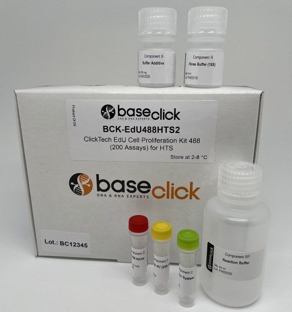
| Dye 488 / 2x 96 well plates | BCK-EdU488HTS2 | € 380,00 |
| Dye 488 / 4x 96 well plates | BCK-EdU488HTS4 | € 590,00 |
| Dye 555 / 2x 96 well plates | BCK-EdU555HTS2 | € 380,00 |
| Dye 555 / 4x 96 well plates | BCK-EdU555HTS4 | € 590,00 |
Chemical Properties
-
Shelf Life
12 months unopened after receipt
-
Storage Conditions
2-8 °C
-
Physical State
kit system made of different components
-
CAS Number
n.a.
-
Excitation (max)
Dye 488: 496 nm | Dye 555: 546 nm
-
Emission (max)
Dye 488: 516 nm | Dye 555: 579 nm
-
Ɛ (max)
Dye 488: 83.000 cm-1M-1 | Dye 555: 91.000 cm-1M-1
-
Preparation/Handling
please see user manual of the kit
Product Information
High-throughput DNA synthesis detection with minimal effort and maximal sensitivity
Our ClickTech EdU Cell Proliferation Kit for High-Throughput Screening (HTS) enables rapid, sensitive and scalable detection of cell proliferation in a 96-well format, ideal for drug discovery, toxicity profiling and compound library screening.
This assay uses the thymidine analogue EdU (5-ethynyl-2′-deoxyuridine), incorporated into DNA during the S-phase of the cell cycle. Detection is achieved via bioorthogonal ‘click’ chemistry, which fluorescently tags EdU-labeled DNA without harsh denaturation steps, delivering bright, stable signals with minimal background, perfect for high-content analysis across diverse cell types [1].

How does the ClickTech EdU assay work?
5-ethynyl-2′-deoxyuridine (5-EdU) is a cell-permeable derivative of thymidine in which the methyl group has been replaced by an ethynyl group. EdU is converted intracellularly to the triphosphate 5-EdUTP and incorporated into newly synthesized DNA during the S phase of the cell cycle, replacing thymidine. The covalent linking of azide-modified fluorophores to EdU via bioorthogonal copper-catalyzed azide–alkyne cycloaddition (CuAAC ‘click’ chemistry) [2, 3] results in the highly specific nuclear labeling of proliferating cells with excellent spatial resolution.

Why is the ClickTech HTS technology superior to other cell proliferation assays?
- Exceptional sensitivity with low background
Click chemistry-based dye conjugation ensures a high signal-to-noise ratio, even at low cell density or under low proliferation conditions.
- Faster, simpler workflow
No DNA denaturation or antibody steps necessary; ready for automation.
- No harsh treatments or specialized equipment needed
Cell morphology and antigenicity are preserved — ideal for multiplexing with immunostaining.
- Scalable and reproducible
Bioorthogonal ‘click’ chemistry ensures consistent assay performance across plates and runs, making it ideal for high-content screening platforms.
Applications
- High-throughput proliferation and cell-cycle screening
- Genotoxicity and mechanism-of-action studies
- Anticancer compound profiling and SAR workflows
- Stem cell proliferation and differentiation monitoring
- Integration with viability/apoptosis markers and HCS imaging
Optimized for High-Content Screening
- Tuned for liquid handling robots and automated plate washers
- Supports thousands of wells with consistent performance
- Unattended batch processing with reproducible results across plates, days, and operators
- Seamless integration with LIMS and data analysis pipelines
LITERATURE
- Multiplexed and reproducible high content screening of live and fixed cells using Dye Drop, C. E. Mills, K. Subramanian, M. Hafner, M. Niepel, L. Gerosa, M. Chung, C. Victor, Benjamin Gaudio, C. Yapp, A. J. Nirmal, N. Clark, P. K. Sorger, 2022, Nat. Commun., 13:6918
- Peptidotriazoles on Solid Phase: [1,2,3]-Triazoles by Regiospecific Copper(I)-Catalyzed 1,3-Dipolar Cycloadditions of Terminal Alkynes to Azides. C. W. Tornøe, C. Christensen, M. Meldal, 2002, J. Org. Chem., Vol. 67, p. 3057-3064.
- A Stepwise Huisgen Cycloaddition Process: Copper(I)-Catalyzed Regioselective “Ligation” of Azides and Terminal Alkynes. V. V. Rostovtsev, L. G. Green, V. V. Fokin, K. B. Sharpless, 2002, Angew. Chemie Int. Ed., Vol. 41, p. 2596–2599.
- Establishment of long-term ostracod epidermal culture, S. Morgan et al., 2020, In Vitro Cell. Dev. Biol. Anim., Vol. 56, p. 760-772. https://doi.org/10.1007/s11626-020-00508-8
- Exposure of vital cells to necrotic cell lysates induce the IRE1α branch of the unfolded protein response and cell proliferation, P. Rohne et al., 2017, Cell Stress and Chaperones, Vol. 23, p. 77-89. https://doi.org/10.1007/s12192-017-0825-6
- Satellite DNA in Vicia faba is characterized by remarkable diversity in its sequence composition, association with centromeres, and replication timing, L. Robledillo et al., 2018, Scientific Reports, Vol. 8, p. 5838. https://doi.org/10.1038/s41598-018-24196-3
- LINC00319 acts as a microRNA-335–5p sponge to accelerate tumor growth and metastasis in gastric cancer by upregulating ADCY3, J. Zou et al., 2019, American Journal of Physiology-Gastrointestinal and Liver Physiology, Vol. 318(1), G10-G22. https://doi.org/10.1152/ajpgi.00405.2018
FAQ
-
What type of cells can incorporate EdU?
Broad compatibility: from bacterial cells (e.g., E. coli) to mammalian cell lines (HeLa, HEK), and even whole organisms (mice, zebrafish, plants).
-
Can I perform EdU cell proliferation detection on living cells?
EdU can be incorporated into living cells, but the detection reaction must be performed on fixed and permeabilized samples.
-
How does EdU labeling compare to BrdU or the 3H-thymidine incorporation assay?
All three methods enable to determine cell proliferation directly by incorporation of a metabolite analogue and subsequent detection. The 3H-thymidine incorporation assay is very sensitive, but the radioactive compound requires specialized equipment and dedicated lab space for handling. EdU and BrdU assays are non-radioactive alternatives with decreased risk for health and environment. Compared to the BrdU incorporation assay the EdU assay is higher sensitivity, faster workflows and avoid harsh DNA denaturing, making it ideal for multiplexing.
-
Can I combine DAPI staining and EdU detection?
Yes, this is feasible. Please note that DAPI staining should be done after the click detection step. Alternatively, SYBR Green DNA staining can be used. But, please note that SYBR green should not be used with dyes of 488 nm wavelengths.
-
When can I safely interrupt the experiment?
It is possible to safely interrupt the protocol after the fixation step. Thereto, remove the fixation solution and wash as suggested by the user manual, then the cells can be stored in buffer at 4° C. Alternatively, the experiment can also be safely interrupted after permeabilization, again as described above.
Please note: It is important to proceed with the experiment if the click cocktail for the detection of the EdU has been prepared already. -
Is antibody staining compatible with the EdU?
Antibody staining is compatible with EdU cell proliferation detection when antibody detection is done after the click detection step. Please be aware of the dye used for EdU detection. Check also the user manual for more information.
-
How to determine the EdU incubation time?
The EdU incubation time depends on the cell type or organism, the applied EdU concentration and the experimental design. For a start it is advisable to refer to a literature protocol (which is close to your experimental setup) and to test the conditions with a low number of samples. As a general guideline we recommend to use a maximum of 10 µM final EdU in the cell culture medium for incubations. For longer incubation (> 1 day) the concentration should be decreased to 1-5 µM.